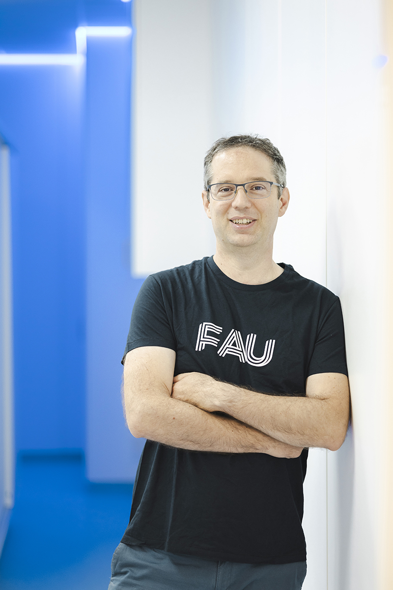
Armin Nagel is developing a new MRI procedure: The biosignature imaging measures the salt content in tissue and can detect changes earlier than is the case to date.
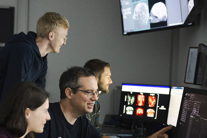
When Armin Nagel looks at MRI images for medical diagnostic purposes, he looks for different details than usually is the case. He does not focus on the structure of pathogenic changes to organs or tissues depicted in cross-sectional images. Instead, the FAU scientist uses the MRI scanner to measure the salt content in tissues. Nagel, who has been Professor of Metabolic and Functional MR Imaging at FAU since 2016, explains, “these ion concentrations often change during the early stages of a disease, even before you can perceive any structural changes.”
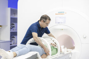
Armin Nagel views ion imaging as a promising approach for diagnosing and treating illnesses, and that is his motivation behind adding this new procedure to the standard repertoire of imaging procedures. Together with his team, he is developing new measuring techniques and evaluation routines, as he explains, “we are trying to see whether we can obtain the images we want that will let us quantify the concentration of sodium or potassium.” The measuring techniques invented by the team are tested on the MR system, initially with phantoms containing, for example, solutions with defined sodium or potassium concentrations. The next step involves testing the method on healthy people, before further investigations are conducted during clinical studies.
Additional data from biosignature imaging
Together with his colleagues Frederik Laun and Moritz Zaiss, Armin Nagel is pursuing the goal of establishing magnetic resonance biosignature imaging. This cutting-edge technological and clinical research focusing on the creation of different image contrasts in the MRI is hoped to provide additional data and new diagnostic findings. In three projects, the researchers are developing rapid MRI techniques capable of characterizing different tissues and their chemical composition and microstructures. While Armin Nagel concentrates on the sub-project ion imaging, Frederik Laun focuses on diffusion-weighted imaging and MRI susceptibility imaging. Moritz Zaiss conducts research into CEST imaging (see information box for further details).
The technological basis for all three projects is 7 tesla magnetic resonance imaging. “Thanks to its increased spectral resolution and the stronger signal, 7 tesla MRI often provides a greater contrast compared to imaging procedures used to date, allowing changes in tissue to be depicted at an earlier stage,” explains Armin Nagel. In the last ten years, the search for clinical applications for 7 tesla MRI has concentrated predominantly on anatomic imaging. Nagel and his colleagues now hope to change that.
The DFG research group “Fast mapping of quantitative and metabolic MRI-fingerprints in ultra-high magnetic field” has received funding of approximately 3.6 million euros from the German Research Foundation to explore this area in greater depth. At FAU, research groups in the area of data science and machine learning led by Katharina Breininger, Florian Knoll and Andreas Maier are working closely together with researchers from Uniklinikum Erlangen. Armin Nagel is the speaker of the DFG research group.
“Ideally, magnetic resonance biosignature imaging will allow changes in the progression of the disease to be detected at an earlier stage and will lead to more effective treatments for diseases.” “
Prof. Dr. Armin Nagel
Detect Alzheimer’s or breast cancer at an earlier stage
MRI biosignature imaging will be used in several research areas in future: In neurodegenerative diseases such as Parkinson’s or Alzheimer’s or in chronic diseases such as chronic kidney disease. The procedure has also sparked interest among those working in oncology. It is hoped that biosignature imaging will lay the foundation for being able to assess individual risk for breast cancer more accurately in future. In all three clinical research fields, the three physicists collaborate with the medical specialists Arnd Dörfler, Jürgen Winkler, Anke Dahlmann, Sabine Ohlmeyer and Sebastian Bickelhaupt from Uniklinikum Erlangen.
“Frederik Laun, Moritz Zaiss and I have known each other for a long time already and we worked together at the German Cancer Research Center in Heidelberg before all three of us accepted a professorship at FAU,” explains Armin Nagel, who before coming to Erlangen was Professor of Experimental Radiology at Universitätsklinikum Ulm and before that the head of a working group at the German Cancer Research Center in Heidelberg. “At FAU, we got together to brainstorm how to merge our research areas to create as much added value as possible in the field of imaging and its applications for diagnostic purposes.”
The potential is huge. “Ideally, magnetic resonance biosignature imaging will allow changes in the progression of the disease to be detected at an earlier stage and will lead to more effective treatments for diseases,” emphasizes Armin Nagel. That is also why the topic sparks his passion: “I find using methods from physics to tackle medical issues fascinating, particularly the diverse opportunities offered by being able to capture non-destructive MRI images from inside the human body.”
MRI biosignature imaging
Within the context of the DFG research group, the researchers Armin Nagel, Frederik Laun and Moritz Zaiss are developing rapid MRI techniques aimed at characterizing various types of tissue, their chemical composition and their microstructures. Armin Nagel focuses on ion imaging (see main text). Frederik Laun concentrates on diffusion-weighted imaging (analysis of water molecule mobility and tissue integrity) and MRI susceptibility imaging (changes in magnetic susceptibility indirectly give indications of the spatial distribution of iron, myelin or calcium levels in the brain, which may be altered in neurodegenerative diseases such as Parkinson’s. Moritz Zaiss focuses on CEST imaging (chemical exchange saturation transfer). These molecular MRI techniques can depict information about proteins and metabolic products which has proven to be useful especially in relation to neurological issues. All three areas of specialization come under the umbrella of magnetic resonance biosignature imaging.
Author: Michael Kniess
This article is part of the FAU magazine
 Innovation, diversity and passion: Those are the three guiding principles of our FAU, as stated in our mission statement. At FAU, we live these guiding principles every day in all that we do – in research, in teaching and when it comes to sharing the knowledge created at our university with society.
This, the second issue of our FAU magazine, underlines all of the above: It shows researchers who tirelessly keep pushing the boundaries of what has been believed to be possible. It introduces students who work together to achieve outstanding results for their FAU, talks about teaching staff who pass on their knowledge with infectious enthusiasm and creativity. And it reports back on members of staff with foresight and a talent for getting to the crux of the matter who are dedicated to improving the (research) infrastructure at FAU as well as people in key positions who are there for their university and are committed to its research location.
Download: FAU Magazin (PDF) Read more articles online