Light in, sound out
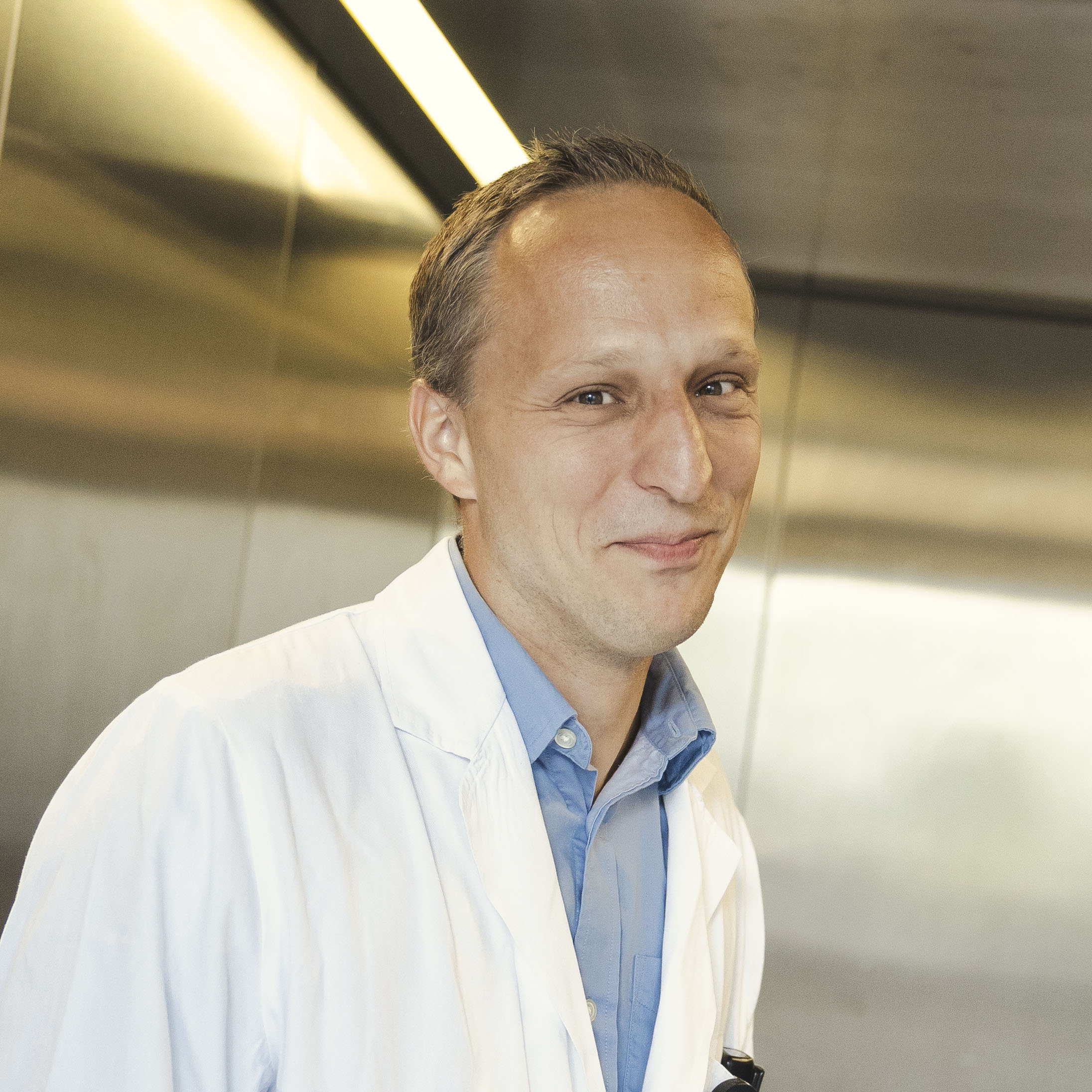
When children come to the Uniklinikum Erlangen complaining of a sore stomach, the search for the cause can prove to be rather challenging. Funded with an ERC Starting Grant, Ferdinand Knieling is conducting research into a new, minimally invasive diagnostic method based on ultrasound. A flying visit to the Department of Pediatrics and Adolescent Medicine.
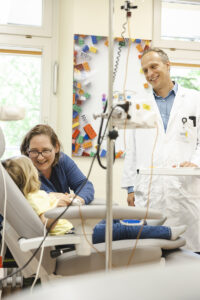
When Ferdinand Knieling strolls through the colorful corridors of the Department of Pediatrics and Adolescent Medicine, he often bumps into parents he’s known for years. “Hello, how are doing?” the senior pediatric physician asks a young family who has come for a regular check-up appointment. “We’re very well, thank you. Our son is really growing and thriving,” the mother smiles, pointing to her son who is hopping along happily beside her. “As a pediatrician, you get so much back,” the physician states later, and explains, “At Uniklinikum we usually deal with chronic and rare diseases that often persist over a long period of time.”
Finding the cause of the diseases is always a challenge and vital for the correct treatment, which is why Knieling has decided to focus on diagnostic research. The fact that he himself is a father of four helps him deal with the children and their parents, who sometimes spend days or weeks at a time in the hospital – at their bedside, in special guest rooms or in the Ronald McDonald house. “The Department of Pediatrics and Adolescent Medicine houses all medical disciplines under one roof: from pediatric surgery and pediatric urology to neonatology, the Pediatric Kidney Center and the rheumatism outpatient clinic, all together between 80 and 100 doctors,” Knieling explains. The patients range in age from premature babies to young adults. “If required, we will still treat a 19 year old if she has multiple pre-existing medical conditions from childhood.”
Ultrasound replaces endoscopy
Ferdinand Knieling’s job involves working on the wards and holding lectures on pediatric medicine for medical students in the Department’s own lecture hall. His third area of responsibility and a project that is close to his heart is research into the early detection of gastrointestinal diseases using special diagnostic ultrasound. “It is not always appendicitis when a child comes to us with a sore stomach,” Knieling explains. “We also observe chronic intestinal diseases such as Morbus Crohn or ulcerative colitis in very young children.” If left untreated, these diseases can trigger inflammation in the whole body and delay growth or the onset of puberty.
Usually, diagnosis requires an invasive gastroscopy or colonoscopy even in young children. However, doctors would prefer to avoid this as far as possible. “It is difficult to carry out an endoscopy on a two year old,” explains the doctor. Even just cleansing the intestines in preparation for the examination by drinking a special liquid can be a torture for the child, so they have to be admitted to the hospital in order to prepare for the endoscopy. That is both time-consuming and costly. Using imaging to make a diagnosis would be less complicated and only minimally invasive.
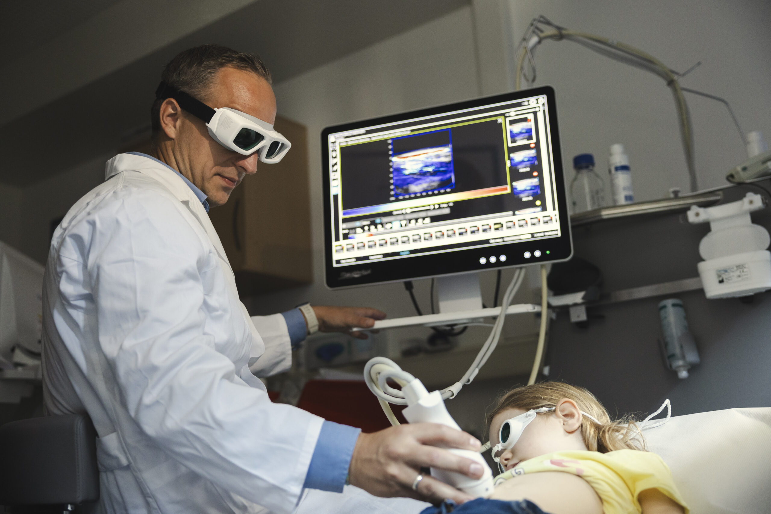
Ferdinand Knieling enters a room where the lights are dimmed and several ultrasound machines are located. The doctor points to a futuristic looking machine used for the procedure known as multi-spectral optoacoustic tomography (MSOT). MSOT is an innovative method that uses short-wave laser light to generate vibrations in the body. Highly sensitive detectors detect these vibrations and use them to create an image, similarly to standard ultrasound. “Light in, sound out: The different substances in the body such as lipids, hemoglobin or connective tissue absorb the light in different ways and become visible. The intestines become even more visible if the patient is given a harmless contrast dye that doesn’t need to be injected and can be swallowed instead,” he explains. “Using improved and more specific dyes in future may allow inflammation in the gut to be pinpointed even more accurately. This method would be particularly well suited to children, as the organs are located only a few centimeters below the surface of the skin and any alterations can be spotted well using the laser method.”
Caterpillar as model of the intestines
The physician and his team have received a Starting Grant from the European Research Council amounting to 1.4 million euros in order to continue to develop the MSOT method over the coming five years. FAU is a worldwide pioneer for this diagnostic method. However, many obstacles have to be overcome before it can be used in regular pediatric practice. “The problem is that medical devices are only authorized for use in patients over the age of 18. The law focuses predominantly on patient safety,” explains project coordinator Knieling. The MSOT method may only be used routinely on children after preliminary studies have been carried out and ethics committees and authorities have conducted the relevant checks.
For this reason, the Erlangen team of researchers are currently conducting experiments on a type of caterpillar. “We have no other choice, we have to be able to provide precise proof that the method is efficient,” says Knieling en route to the laboratory on the third floor in the clinic. The caterpillars are dissected carefully and prepared for the laser ultrasound. The images are then analyzed in a computer room. “We are working with an interdisciplinary team of biologists, engineers and computer scientists.” The pediatrician hopes that the minimally invasive ultrasound method will one day become the standard method for small patients, in order to be able to provide them the treatment they need quickly and accurately.
Preliminary studies with moth larvae
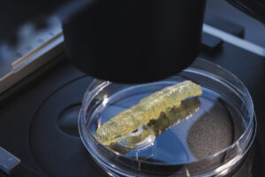
Before the MSOT method can become the standard procedure for pediatric diagnostics, a number of preliminary studies are required. With this aim in mind, Ferdinand Knieling’s team is conducting research together with Dr. Anton Windfelder from Uniklinikum Gießen on the larvae of the tobacco hornworm, a moth. The invertebrate is especially suited as a model organism for preclinical studies on chronic inflammatory bowel diseases, as up to 75 percent of the genes that can trigger a disease in humans are shared by these insects. Unlike the case with animal experiments on mice, there is no complicated approval procedure to comply with when it comes to larvae. Other advantages of using larvae rather than rats or mice are they reproduce more rapidly, are less expensive to keep and entail fewer ethical concerns. At the Department of Pediatrics and Adolescent Medicine, the caterpillars are prepared using a special chemical solution until they become transparent. In this way, researchers can then prove that the new dyes have indeed reached their target in the intestines.
Author: Susanne Stemmler
This article is part of the FAU magazine
 Innovation, diversity and passion: Those are the three guiding principles of our FAU, as stated in our mission statement. At FAU, we live these guiding principles every day in all that we do – in research, in teaching and when it comes to sharing the knowledge created at our university with society.
This, the second issue of our FAU magazine, underlines all of the above: It shows researchers who tirelessly keep pushing the boundaries of what has been believed to be possible. It introduces students who work together to achieve outstanding results for their FAU, talks about teaching staff who pass on their knowledge with infectious enthusiasm and creativity. And it reports back on members of staff with foresight and a talent for getting to the crux of the matter who are dedicated to improving the (research) infrastructure at FAU as well as people in key positions who are there for their university and are committed to its research location.
Download: FAU Magazin (PDF) Read more articles online