Using “tattoos” to make damaged muscle tissue visible
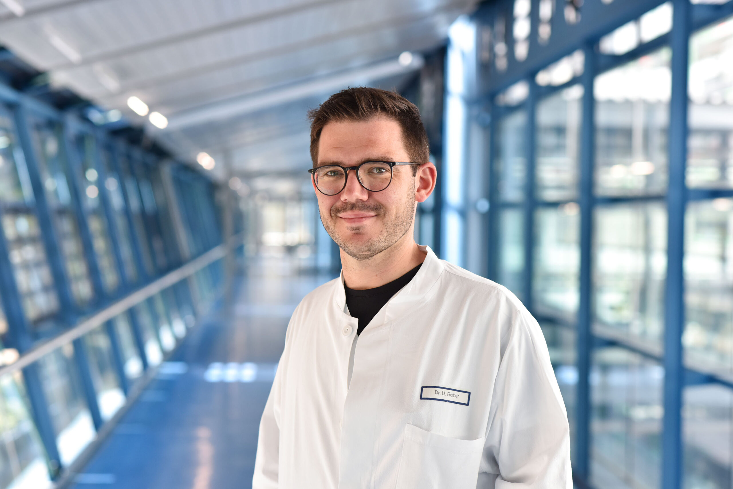
Arterial occlusion in the legs mainly affects older people and can often require surgery to treat it. A research team led by PD Dr. Ulrich Rother, deputy head of the Department of Vascular Surgery (head: Prof. Dr. Werner Lang) at Uniklinikum Erlangen, is currently developing new and non-invasive diagnostic methods in order to improve treatment of peripheral artery disease (PAD). “We are using a new method called multispectral optoacoustic tomography (MSOT), which allows us to monitor biological processes in the body in real time without radiation”, explains Dr. Rother.
Foundation provides more than 300,000 euros of funding for innovative diagnostics of arterial occlusion
The vascular specialist is researching the method in an interdisciplinary team at Uniklinikum Erlangen with Dr. Alexander Seitel and Prof. Dr. Lena Maier-Hein from the German Cancer Research Center in Heidelberg. The team aim to map the blood flow and oxygen supply to muscle tissue non-invasively with three-dimensional imaging. The researchers in Erlangen and Heidelberg have now received a total of 318,168 euros of funding from the Else Kröner-Fresenius-Stiftung for their joint clinical study.
There has not been a comparable method for diagnosing muscle conditions up to now. Multispectral optoacoustic tomography is a safe and non-invasive method used to measure the composition and oxygen supply of muscle tissue. “We use the optoacoustic effect for this purpose – in short, ‘light in, noise out’”, says Dr. Rother about the innovative method that he is investigating at Uniklinikum Erlangen with PD Dr. Ferdinand Knieling, senior physician at the Department of Pediatrics and Adolescent Medicine (director Prof. Dr. Joachim Wölfle) and Prof. Dr. Maximilian Waldner, senior physician at the Department of Medicine 1– Gastroenterology, Pneumology and Endocrinology (director: Prof. Dr. Markus F. Neurath). “To allow three-dimensional imaging of the vascular system being investigated, we have developed an optical pattern especially for the longitudinal scan which is printed on film using special ink and then applied to the skin,” says Dr. Seitel, who is part of the team in Heidelberg. Like a self-adhesive tattoo, this film is applied to the tissue to be examined and a high-resolution three-dimensional image is produced using MSOT. This method, known informally as “tattoo tomography”, was developed at the German Cancer Research Center. “For future diagnostics, we want to use the potential of this tattoo tomography in our study, which is easy to use, to gain detailed insights into the oxygen supply and damage to the muscle tissue being examined,” explains Ulrich Rother. “This new method would allow physicians to diagnose peripheral artery disease, which is on the rise worldwide, even more accurately,” explains vascular surgeon Rother.
Else Kröner-Fresenius-Stiftung
The charitable foundation Else Kröner-Fresenius-Stiftung, is dedicated to funding medical research and supports medical and humanitarian projects. The foundation was established in 1983 by entrepreneur Else Kröner and made into her sole heir. It has provided funding to around 2,600 projects to date. With a current annual funding volume of around 70 million euros, it is one of the largest foundations that supports the field of medicine in Germany.
Further information
PD Dr. Ulrich Rother
Phone: +49 9131 85 32968
gefaesschirurgie-sekr@uk-erlangen.de